Your body fought off the virus — but damaged your lungs
Researchers zero in on potential key to rapidly repairing tissue harmed by inflammation
Clea Simon
Harvard Correspondent
4 min read
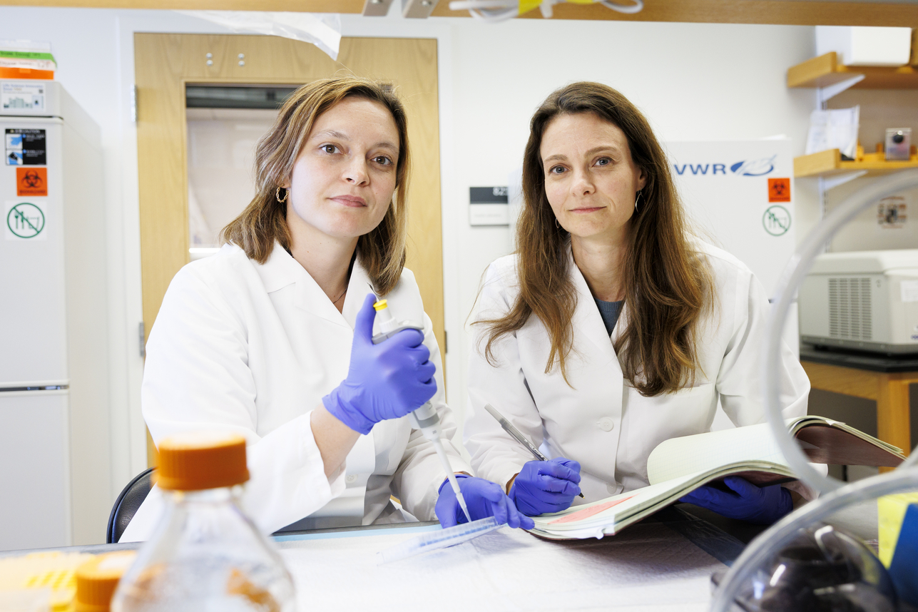
Daisy Hoagland (left) and Ruth Franklin.
Photos by Niles Singer/Harvard Staff Photographer
When the lungs are attacked by a virus, the damage doesn’t stop there. The body’s natural defenses cause inflammation while fighting the virus, often leaving lasting problems. The cells that make up the lungs’ mucosal lining are exposed to the environment with every breath — both highlighting the risk of infection and emphasizing the need for a robust response. In a paper published recently in Science, a team of Harvard researchers reveals how macrophages — a type of immune cell — may be key to repairing that damage, both from the initial infection and from the immune response itself.
“Nearly every tissue in the body contains resident macrophages,” explained Ruth Franklin, assistant professor of stem cell and regenerative biology, in whose lab the research was conducted. These multifunctional cells “help to maintain function of tissues.”
The problems arise when the lungs, which regularly encounter viruses and bacteria, become infected. The organ’s first priority is to fight the infection. However, the body’s reaction can damage the epithelial cells that line organs and form their protective barriers.
“What we found is that macrophages in the lung produce a growth factor, oncostatin M (or OSM), that is able to quickly restore the epithelial barrier in the lung,” Franklin said. “This rapid repair is extremely important because in the lung, you’re more vulnerable to the outside environment if you don’t have that barrier.”
“What we found is that macrophages in the lung produce a growth factor, oncostatin M (or OSM), that is able to quickly restore the epithelial barrier in the lung.”
Ruth Franklin
Daisy Hoagland, a postdoctoral researcher in Franklin’s lab and co-first author of the paper, elaborated: “There are different kinds of viruses, but some viruses go into a cell, hijack all of its machinery, and cause the cell to rupture. Also, the virus can cause the cell to self-destruct intentionally.
“A lot of the damage will happen because the immune system is trying to kill the infected cells,” she added. “It’s really hard to repair the epithelial barrier during inflammation because a lot of the inflammatory signals prevent cells from replicating and redirect cells toward defense programs instead of regeneration.”
To test whether OSM was important for repairing the epithelial barrier, the team studied mice that had been bioengineered to prevent production of OSM and infected them with the influenza virus. “By nearly every metric that we checked, mice lacking OSM had more damage than normal mice,” Hoagland said.
The next step involved using a synthetic virus-like molecule, poly(I:C), which doesn’t replicate like a real virus but nonetheless triggers an immune response. Importantly, it activates the same inflammatory signals that usually prevent cells from dividing. Using this on both the OSM-deficient mice and normal mice led to the same results. The conclusion? OSM is essential to helping the lung’s protective lining heal during the immune system’s antiviral response.
“Cells die in response to both viral infection and from the inflammation it causes,” said Franklin. “Repairing this damage is difficult while an infection is ongoing, but OSM can override some of these signals and restore the barrier.”
“A lot of the time people who die on ventilators from diseases like COVID-19 or severe viral pneumonia have actually cleared the virus, but they can’t fix their lungs in the context of all of this inflammation.”
Daisy Hoagland
This latest discovery follows years of studies of OSM that began around 2014, noted Franklin — “before I came to Harvard.” It also opens new paths of exploration. For example, said Hoagland, researchers do not yet know what OSM does when there is no infection.
“What we found is that OSM is produced at low levels in the absence of inflammation,” she said. “We’re currently trying to understand the role of OSM at baseline.”
In the meantime, the team’s research continues.
“Right now, we’re trying to see if there’s any therapeutic potential for OSM,” Hoagland said. Although the experimentation continues in mice, the goal is to see whether OSM could help repair human lungs damaged by illness.
“A lot of the time people who die on ventilators from diseases like COVID-19 or severe viral pneumonia have actually cleared the virus, but they can’t fix their lungs in the context of all of this inflammation,” Hoagland said. “We’re really hopeful that OSM could potentially be therapeutically beneficial in those situations.”
This work was funded in part by the National Institutes of Health and the National Science Foundation.